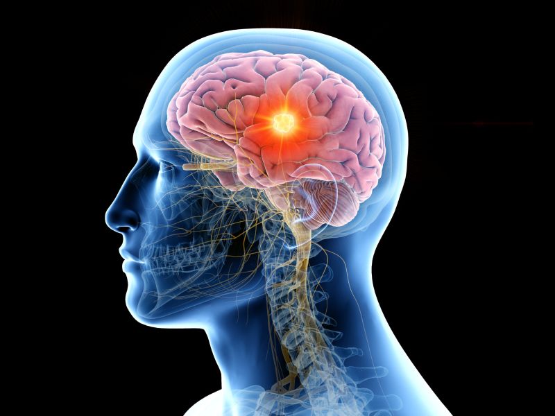
Key neural systems in the brain that regulate movement initiation have been discovered by researchers. The brain’s mesencephalic locomotor region (MLR), an ancient region that has survived in all vertebrates, is essential for the initiation of movements including walking, flying, and swimming.
The study made use of the zebrafish larvae’s transparent brains to examine and map the neural circuits that initiate forward movement. This finding holds great promise for understanding and treating motor deficits, including those seen in people with Parkinson’s disease.
Important facts:
From walking to swimming, the mesencephalic locomotor region (MLR) in the brain is essential for starting a variety of actions in different animals.
Researchers created a new technique that tracks nerve impulse propagation in the brain’s locomotor structures by using the transparency of zebrafish larvae brains.
The study discovered that zebrafish forward movement vigour and MLR stimulation intensity are correlated, potentially providing new information about gait transitions in both aquatic and terrestrial animals.
“These neurons control the finer details of movement, such as starting, stopping, and changing direction. In a way, they give steering instructions! Previous work on mice had revealed that reticulospinal neurons control turning; Martin and Mathilde have discovered the control circuit that triggers forward locomotion”, Claire Wyart says.
“Animals move to explore their environment in search of food, interaction with others, or simply out of curiosity. But the perception of danger or a painful stimulus can also activate an automatic flight reflex”, Post-doctoral fellow at the Paris Brain Institute Martin Carbo-Tano explains.
Movement start in both situations is dependent on the activation of so-called reticulospinal control neurons, which are interconnected in the brainstem, the most posterior region of the brain.
These neurons are crucial for motor control of the limbs and trunk as well as movement coordination since they transfer nerve impulses between the brain and spinal cord.
The mesencephalic locomotor region (MLR), which is located above the reticulospinal neurons, is also crucial for locomotion since, in animals, its stimulation initiates forward propulsion. Numerous creatures, including guinea pigs, cats, salamanders, and even lampreys, exhibit it.
“Because the role of the MLR is conserved in many vertebrate species, we assume that it is an ancient region in their evolution—essential for initiating walking, running, flying, or swimming,” he adds.
“But until now, we didn’t know how this region transmits information to the reticulospinal neurons. This prevented us from gaining a global view of the mechanisms that enable the vertebrae to set themselves in motion and, therefore, from pointing out possible anomalies in this fascinating machinery”.
The control tower unlocks its doors.
It can be challenging to study movement initiation because neurons in the brain stem are difficult to access and difficult to observe in vivo in a moving animal. Martin Carbo-Tano has created a novel method to activate microscopic brain regions in order to address this issue.
The researchers used the transparency of the zebrafish larvae brain in collaboration with Mathilde Lapoix, a Ph.D. student on Claire Wyart’s team at the Paris Brain Institute, to localise the locomotion-related regions downstream of the MLR and track the transmission of nerve impulses.
This approach, which was influenced by the research of their associate Réjean Dubuc at the University of Montréal, allowed them to produce a number of amazing findings.
“We observed that neurons in the mesencephalic locomotor region are stimulated when the animal moves spontaneously, but also in response to a visual stimulus. They project through the pons—the central part of the brain stem—and the medulla to activate a subpopulation of reticulospinal neurons called ‘V2a’.
The midbrain is a region of intense focus.
The researchers stimulated the mesencephalic locomotor area in an effort to experimentally trigger this mechanism in order to better understand how it affects the movements of larval zebrafish. They discovered that the length and force of forward motion were proportional to the level of stimulation.
“Quadrupeds can adopt different gaits, such as walking, trotting, or galloping. But aquatic animals also mark gait transitions”, Martin Carbo-Tano adds.
“We think that MLR has a role to play in this intensification of movement, which we have observed in zebrafish.”
This research allowed for the first time the mapping of the neural circuits responsible for initiating forward movement, a function that is impaired in people with Parkinson’s disease.
This is a crucial step in understanding the motor control processes that occur upstream of the spinal cord. One day, it may be feasible to identify and regulate each reticulospinal neuron individually in order to simulate in detail how locomotion functions and fix any that are malfunctioning.

 Diabetology2 weeks ago
 Diabetology1 week ago
 Diabetology3 days ago
 Diabetology3 days ago
 Diabetology3 days ago
 Diabetology1 day ago


