Pulmonology
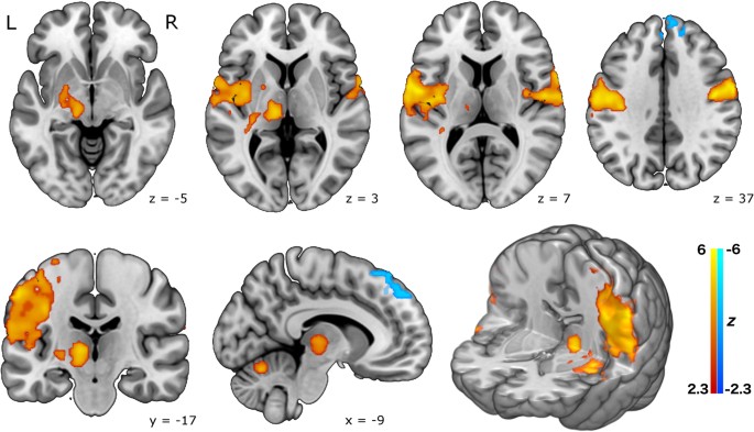
Smoking Severity Affects Brain Gray Matter in Opioid Use Disorder
-

 Diabetology1 week ago
Diabetes Screening: Why Early Detection Saves Lives
-

 Diabetology7 days ago
Glycemic Index Chart + Tips: Best & Worst Foods for Blood Sugar
-

 Diabetology5 days ago
Daily Diabetes Management Plan for Type 2 Diabetes
-

 Diabetology1 week ago
Caring for Diabetes Daily: A Practical Guide to Routine Control
-

 Diabetology1 week ago
Hyperglycemia: What You Need to Know to Stay Safe
-

 Diabetology5 days ago
Understanding the Impact of High Glucose Levels
-

 Diabetology3 days ago
How Real-Time Blood Glucose Monitors Improve Health Outcomes
-

 Diabetology3 days ago
How a Plant-Based Diet Can Help Manage Type 2 Diabetes


