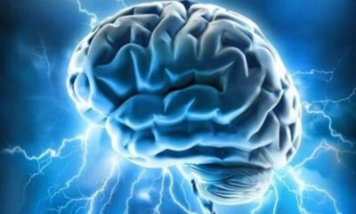
Scientists at Cedars-Sinai have made an important discovery on the complex processes involved in working memory in the human brain. Under the direction of postdoctoral fellow Jonathan Daume at Cedars-Sinai’s Rutishauser Lab, the team has discovered a subset of neurons that are essential for integrating the brain’s concentration and store processes for short-term memory recall.
Working memory requires the careful balancing act of purposeful focus and short-term memory storage. For example, it is necessary to remember a phone number long enough to dial it. This type of memory is especially brittle and necessitates constant attention because it only allows the brain to store information for a few seconds. The study’s senior author and director of Cedars-Sinai’s Center for Neural Science and Medicine, Ueli Rutishauser, highlights how susceptible working memory is, particularly when it comes to conditions like attention-deficit hyperactivity disorder and Alzheimer’s disease.
The research used a novel method to investigate the inner workings of working memory. It recorded the brain activity of 36 hospitalized patients who had electrodes surgically implanted as part of the diagnosis process for epilepsy. The results were published in the esteemed journal Nature. While the patients performed a task requiring working memory, the researchers attentively observed the activity of individual brain cells and brain waves. Either one or three photos were shown to the patients, and then there was a brief period of time when the screen was blank and they had to recall the images they had just seen. They were then asked to identify if the next picture they saw was one of the earlier ones.
Notably, researchers saw the firing of two different groups of neurons when patients completed the working memory task successfully and quickly: “phase-amplitude coupling” (PAC) neurons, a newly discovered group in this study, and “category” neurons, which fired in response to the particular categories shown in the photos. The PAC neurons use a technique known as phase-amplitude coupling to make sure that the category neurons focus and store the learned information, even though they are not in charge of content storage. The PAC neurons improve the working memory recall capacity of the category neurons by timing their activity with theta and gamma waves in the brain.
Rutishauser uses the following analogy to explain this phenomenon: “Imagine that when a patient views a picture of a dog, their category neurons fire “dog, dog, dog,” but the PAC neurons fire “focus/remember.” “Remember dog” is the consequence of the two sets of neurons creating harmony by superimposing their messages through phase-amplitude coupling.
Surprisingly, the PAC neurons perform this vital task in the hippocampus, a part of the brain that is typically linked to long-term memory. This study sheds fresh light on the mechanics underpinning the brain’s memory processes by offering the first evidence that the hippocampus is also involved in working memory regulation.
Led by Cedars-Sinai in partnership with the University of Toronto and the Johns Hopkins School of Medicine, this ground-breaking study was carried out as part of a multi-institutional consortium supported by the National Institutes of Health’s Brain Research Through Advancing Innovative Neurotechnologies Initiative (The BRAIN Initiative). The NIH BRAIN Initiative’s director, Dr. John Ngai, highlights the importance of this research in understanding intricate brain functions, especially in light of debilitating brain illnesses like Alzheimer’s disease and other dementias.
An important step forward in our comprehension of the complex workings of the brain has been made with the identification of the network of neurons that coordinates cognitive control and the storage of sensory information in working memory. This realization offers hope to those suffering from neurological illnesses that impair working memory by potentially leading to the discovery of novel treatments for these crippling conditions.

 Diabetology2 weeks ago
 Diabetology6 days ago
 Diabetology5 days ago
 Diabetology5 days ago
 Diabetology4 days ago
 Diabetology2 days ago
 Diabetology2 days ago
 Diabetology7 hours ago
